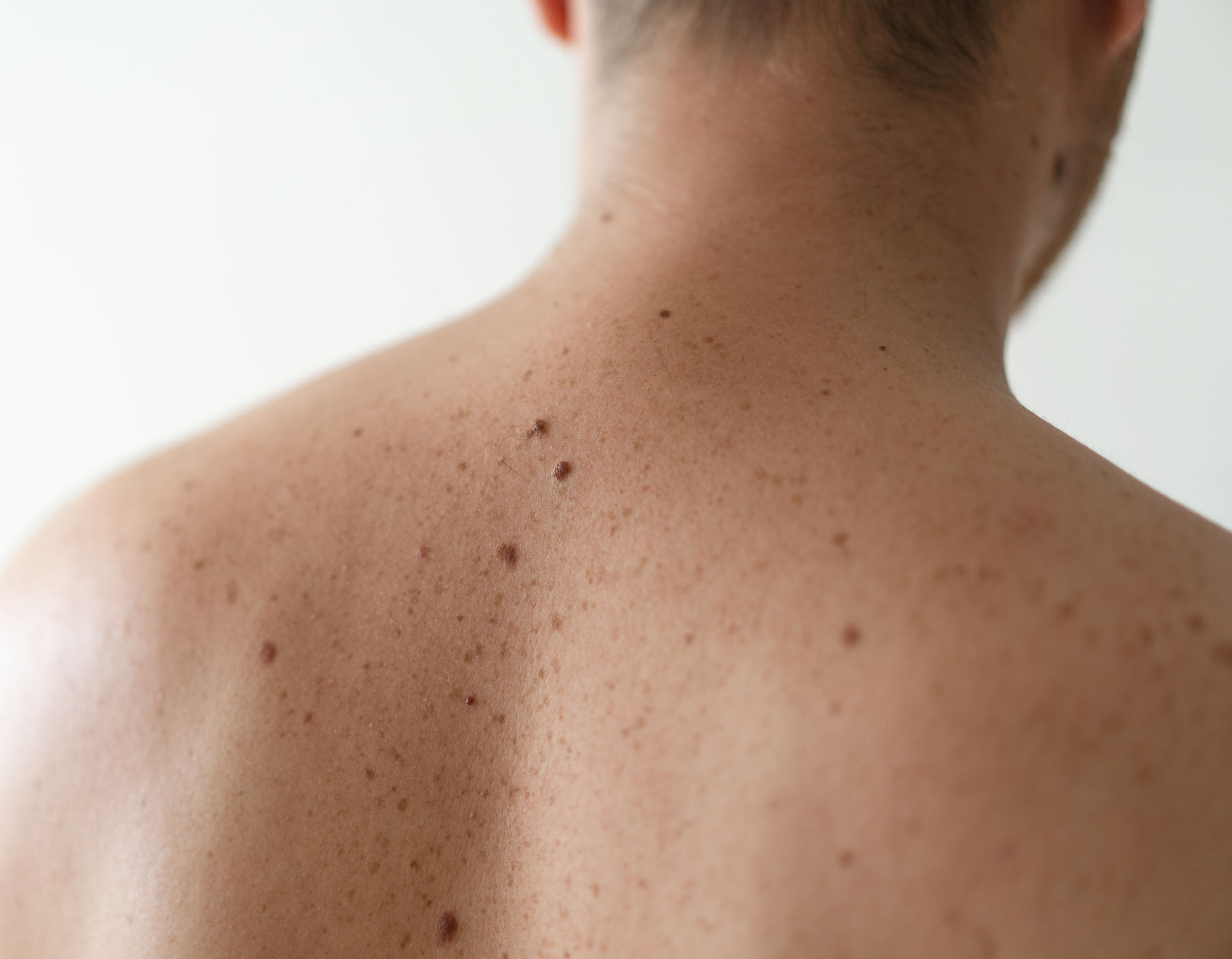
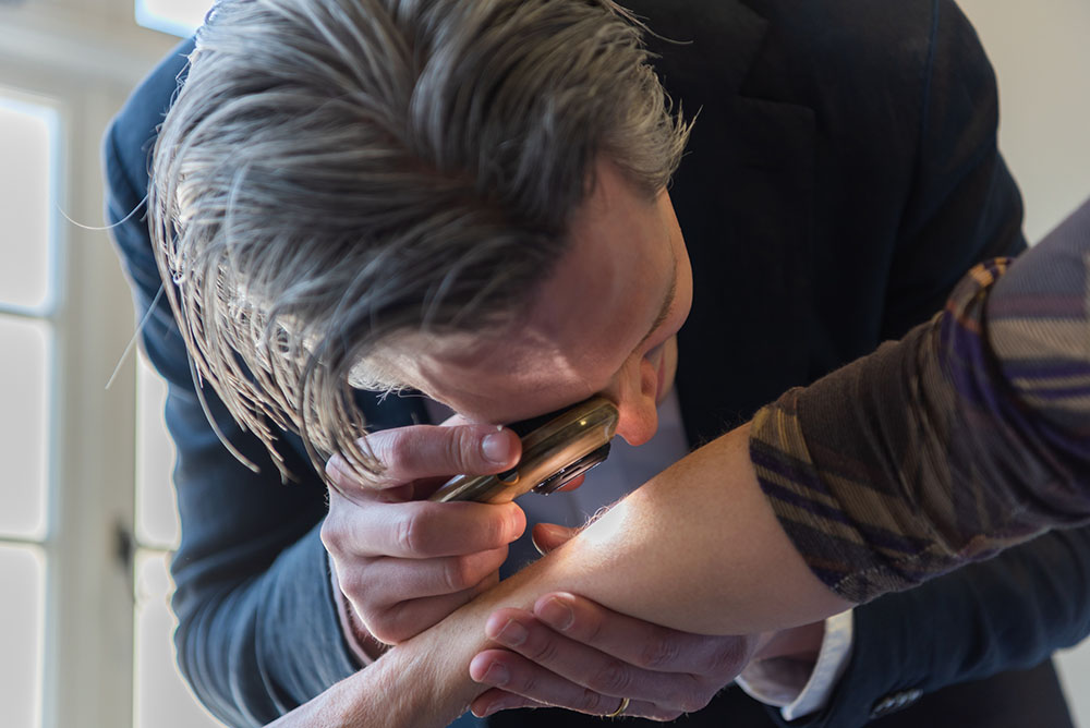
Mole checks are one of the most common reasons why patients will attend the clinic. I have a particular interest in the diagnosis and surgical treatment of skin cancer, I lead the melanoma multidisciplinary team meeting at Guy's and St Thomas' NHS Trust and I have published on the role of AI in skin cancer diagnosis so I am well equipped to efficiently and comprehensively examine your skin and help you to decide on a plan for treatment.

Skin cancer results from unregulated growth of the cell types that make up the skin. The three most common forms of skin cancer are basal cell carcinoma, squamous cell carcinoma and melanoma.
Whilst the term 'mole' is sometimes used to refer to any lesion on the skin the term more accurately refers to growths of pigment cells (melanocytes). Melanocytes are present throughout the skin surface and are responsible for tanning and pigmentation of the skin. The medical name for moles is melanocytic naevi.
Melanoma is the most serious form of skin cancer since it has the potential to spread elsewhere in the body. This risk is much lower if a melanoma is detected at an early stage and this is why it is important to identify melanomas as early as possible. It may arise either within a pre-existing mole or in an area of normal skin. I will carefully examine any moles of concern with a microscope - a dermatoscope. Whilst the diagnosis of melanoma can be apparent to the naked eye, in some cases early melanoma may only be detected through subtle irregularities of the pattern of pigmentation that are seen with the dermatoscope.
Basal cell carcinoma is the most common form of skin cancer. It generally is not dangerous to your overall health, however it can cause extensive local tissue damage if allowed to grow and therefore it is important that it is diagnosed early. Basal cell carcinoma usually has the appearance of a small, shiny lesion on the skin surface. You may notice it because it bleeds or does not heal. As for melanoma, basal cell carcinoma has characteristic appearances when examined with the dermatoscope that helps me to make a confident diagnosis.
Squamous cell carcinoma is less common than basal cell carcinoma, however it is more serious since it does carry a small risk of spreading elsewhere in the body. It usually presents as a fast-growing lump on the skin although occasionally appearances can be more subtle.

Skin cancer can present as a new growth on the skin, a change in the appearance of a mole or a bleeding or ulcerated lesion. In general a new or changing lesion on the skin should be checked.
The following is a simple method that can help to identify moles that may be of concern:
It is important to note that not all moles fulfilling one or more of the above criteria is a melanoma. Conversely, some (usually early) melanomas will not fulfill these criteria and may only be detected on microscope (dermascopic) examination of the mole.
All forms of skin cancer are more common in individuals who have had increased levels of sun exposure or who have used sun beds. Skin cancer is also significantly more common in those with lighter skin types.
Other risk factors for skin cancer include:
During a skin check I am looking firstly for skin cancer, secondly for any precancerous change and finally for any other skin conditions that might be present.
I will begin the consultation by asking you some questions about what you have noticed on the skin - that might be a new mole or change in the appearance of an existing skin lesion. I will then ask you some more general questions about your general health and whether you or any family member has had skin cancer.
I will generally start by checking any skin lesions that you have noticed. If you are female, a female chaperone will be present should you need to undress. You can also request a chaperone if you are male. I will use a dermatoscope - a microscope that allows me to examine skin lesions at high resolution. Different skin lesions exhibit characteristic patterns when viewed with the dermatoscope and this helps me to make an accurate diagnosis.
If you would like me to perform a full skin check, I will perform a systematic examination of your skin - starting from the head and neck and working through all body areas. I will ask your permission to take dermascopic (microscope) photographs of any skin lesions of concern in order that the appearance is recorded in your patient record. If you have a large number of moles I may arrange for you to have mole mapping photographs performed in order that we can monitor for any changes in the future.
If you have a number of irregular moles I may advise that you have mole mapping photographs performed. This enables me to monitor for any change in your moles over time. This is performed with the VECTRA WB360 whole body 3D imaging system - the most advanced 3D skin imaging system in the UK.
The principle of mole mapping is that harmless moles either do not change in appearance or change very slowly over years whereas melanomas usually will change significantly in appearance over a number of months.
Mole mapping consists of photography of the skin surface. It serves as a photographic record of lesions on the skin for comparison in future consultations. The photography itself does not perform any analysis of the skin and does not replace a comprehensive examination of the skin by a dermatologist with a dermatoscope (microscope). For any moles that I am concerned about, I will take additional digital dermascopic photographs of the pigment pattern. This allows me to check for any subtle changes at a follow up appointment.
Even if you are having regular skin checks with a dermatologist it is important that you monitor your own skin at home. I generally advise patients to perform a full skin check every few months - this has the benefit that you learn what is normal on your own skin and learn to recognise any change. An easy way to monitor for change is to take photographs of all of your skin (you will need help for some areas) and then repeat these photographs after a few weeks/months. If you notice any new skin lesions or any change in the appearance (size, shape, color) of existing skin lesions it is important that these are reviewed.
Skin cancer can have the appearance of an irregular mole, a lump on the skin surface or a non-healing area of skin. However, if you have a lot of skin lesions it can be very difficult to know which you should be worried about. In this scenario I would generally advise that you have a skin check with a dermatologist and then subsequently monitor for any change yourself.
If I am concerned about a skin lesion then I may recommend a skin biopsy - either a sample of the lesion or the entire lesion is removed under local anaesthetic for analysis. Depending upon the nature of a skin lesion it can be removed either by cutting and stitching to leave a straight line scar or by shaving parallel with the skin surface and I will discuss the different options with you. The lesion is then sent for analysis by an experienced pathologist with whom I have a close working relationship and results are typically available within 1-2 weeks.
I have advanced training in skin surgery and have performed thousands of skin biopsies so where this is required I can perform this with minimal discomfort and scarring. You can learn more about what to expect during skin surgery here.
To book an in person consultation, enter your details below and my practice management team will contact you to schedule the appointment. Alternatively call 0203 389 6076 (calls are answered during working hours) or email: contact@drmagnuslynch.com.