Understanding Skin Cancer: Prevention, Detection and Treatment


Skin cancer is the most common form of cancer in the UK, and rates are continuing to rise. However, the good news is that many skin cancers are preventable, and when detected early, most are highly treatable. As a Consultant Dermatologist and Dermatological Surgeon with a special interest in skin cancer, I treat many patients with complex skin cancers. This article aims to provide you with a clear understanding of skin cancer – what it is, how you can detect it early, and the treatment options available should you need them.
What is Skin Cancer?
Skin cancer results from unregulated growth of the cell types that make up the skin. This occurs when DNA damage to skin cells – most often caused by ultraviolet (UV) radiation from the sun or tanning beds – triggers mutations. Over time, these mutations allow skin cells to multiply and form tumours.
Whilst the term 'mole' is sometimes used to refer to any lesion on the skin the term more accurately refers to growths of pigment cells (melanocytes). Melanocytes are present throughout the skin surface and are responsible for tanning and pigmentation of the skin. The medical name for moles is melanocytic naevi.
The three most common forms of skin cancer are:
- Basal Cell Carcinoma (BCC): This is the most common form of skin cancer. It generally is not dangerous to your overall health, however it can cause extensive local tissue damage if allowed to grow and therefore it is important that it is diagnosed early. Basal cell carcinoma usually has the appearance of a small, shiny lesion on the skin surface. You may notice it because it bleeds or does not heal. As for melanoma, basal cell carcinoma has characteristic appearances when examined with the dermatoscope that helps me to make a confident diagnosis.
- Squamous Cell Carcinoma (SCC): Squamous cell carcinoma is less common than basal cell carcinoma, however it is more serious since it does carry a modest risk of spreading elsewhere in the body. It usually presents as a fast-growing lump on the skin, a non-healing area on the skin, or a flat lesion with a scaly, crusted surface.
- Melanoma: Melanoma is the most serious form of skin cancer since it has the potential to spread elsewhere in the body. This risk is much lower if a melanoma is detected at an early stage and this is why it is important to identify melanomas as early as possible. It may arise either within a pre-existing mole or in an area of normal skin. I will carefully examine any moles of concern with a skin microscope known as a dermatoscope. Whilst the diagnosis of melanoma can be apparent to the naked eye, in some cases early melanoma may only be detected through subtle irregularities of the pattern of pigmentation that are seen with the dermatoscope.
Risk Factors for Skin Cancer
While anyone can develop skin cancer, certain factors can increase your risk. Understanding these can help you take proactive steps.
Other risk factors for skin cancer include:
- Sun Exposure: This is the leading cause. Cumulative sun exposure throughout your life, as well as episodes of severe sunburn (especially in childhood), significantly increase risk.
- Tanning Beds: Artificial tanning lamps and beds emit UV radiation and are a proven cause of skin cancer. There is no such thing as a 'safe tan' from a sunbed.
- Skin Type: People with fair skin are at higher risk.
- Previous Skin Cancer: Individuals who have previously suffered from melanoma or other forms of skin cancer have a higher risk of developing other skin cancers in the future.
- Family History: A family history of skin cancer, particularly melanoma, increases your risk.
- Number of Moles: Having many moles, or certain types of irregular moles (dysplastic naevi), is a risk factor.
- Weakened Immune System: Conditions or medications that suppress the immune system (e.g., immunosuppressive medications for an organ transplant) can make you more susceptible.
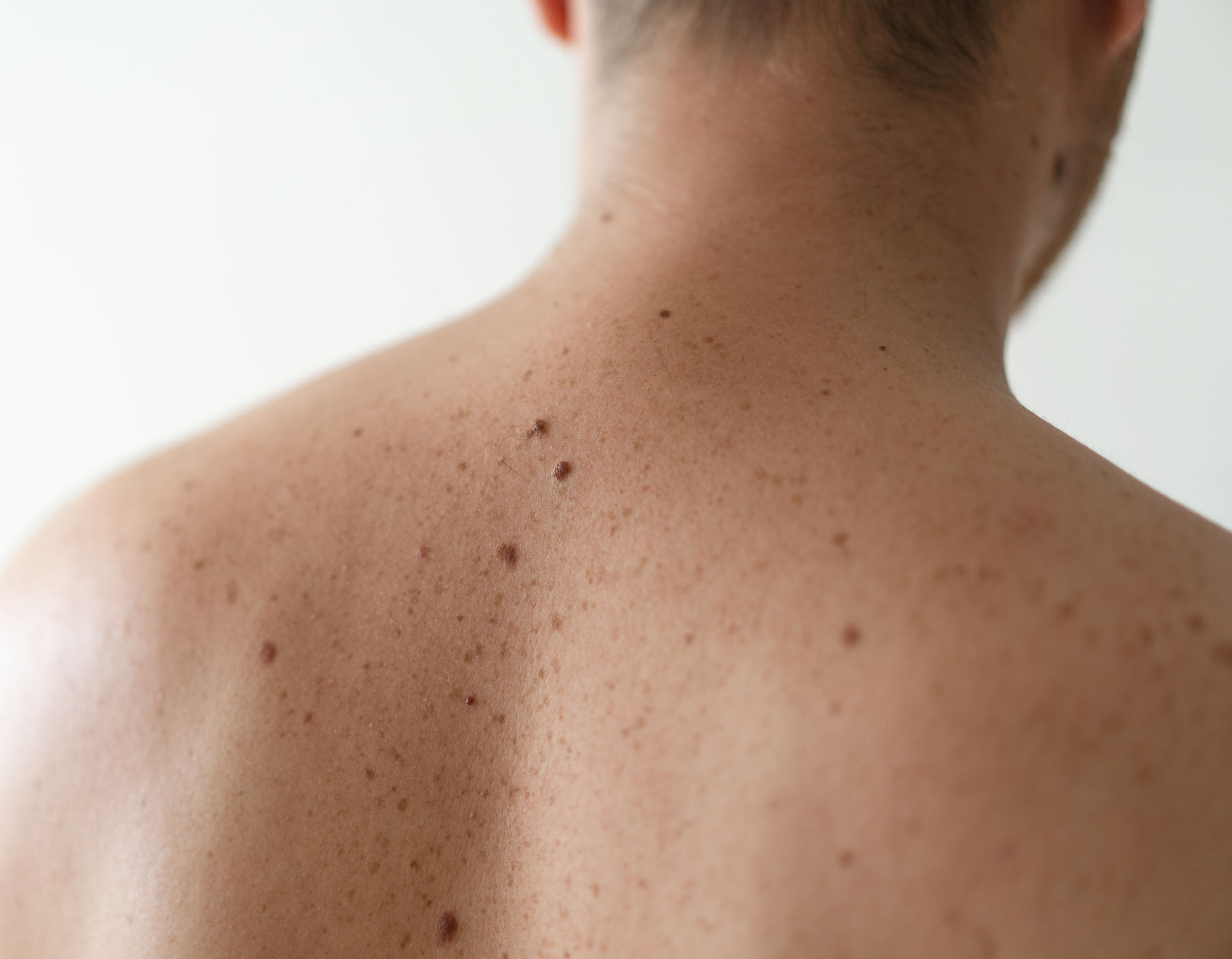
How to Detect Skin Cancer at Home
When it comes to skin cancer, early detection is important. The earlier a skin cancer is identified and treated, the better the outcome. This is where regular self-examinations play a vital role.
What does skin cancer look like?

Skin cancer can present as a new growth on the skin, a change in the appearance of a mole or a bleeding or ulcerated lesion. In general a new or changing lesion on the skin should be checked by a doctor.
The following is a simple method that can help to identify moles that may be of concern (the ABCDE rule):
- Asymmetry: Harmless moles are more often symmetrical (one side is a mirror image of the other) whereas melanoma is more likely to be asymmetrical.
- Border: Harmless moles usually have a smooth, regular border whereas melanomas may have a rough, irregular border.
- Colour: The presence of more than two colors (red, brown, blue, black) is more common in melanoma.
- Diameter: Melanomas are more likely to have a diameter of more than 6mm (about the size of a pencil eraser), although melanomas can sometimes be smaller than this.
- Evolution: Melanomas will usually change noticeably in appearance over a period of several months. This can also include any new symptom such as bleeding, itching or crusting.
It is important to note that not all moles fulfilling one or more of the above criteria is a melanoma. Conversely, some (usually early) melanomas will not fulfill these criteria and may only be detected on microscope (dermatoscopic) examination of the mole.
For non-melanoma skin cancers (BCC and SCC), look for new growths, spots, or lesions that don't heal. If you notice any of these signs, or anything else that concerns you, it's crucial to see a dermatologist.
How to check your skin at home
Even if you are having regular skin checks with a dermatologist it is important that you monitor your own skin at home. I generally advise patients to perform a full skin check every few months - this has the benefit that you learn what is normal on your own skin and learn to recognise any change. An easy way to monitor for change is to take photographs of all of your skin (you will need help for some areas) and then repeat these photographs after a few weeks/months. If you notice any new skin lesions or any change in the appearance (size, shape, color) of existing skin lesions it is important that these are reviewed.
Skin cancer can have the appearance of an irregular mole, a lump on the skin surface or a non-healing area of skin. However, if you have a lot of skin lesions it can be very difficult to know which you should be worried about. In this scenario I would generally advise that you have a skin check with a dermatologist and then subsequently monitor for any change yourself.
What to Expect During a Skin Check
Skin checks are one of the most common reasons why patients will attend the clinic. During a skin check I am looking firstly for skin cancer, secondly for any precancerous change and finally for any other skin conditions that might be present.
I will begin the consultation by asking you some questions about what you have noticed on the skin - that might be a new mole or change in the appearance of an existing skin lesion. I will then ask you some more general questions about your general health and whether you or any family member has had skin cancer.
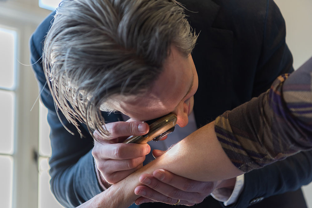
I will generally start by checking any skin lesions that you have noticed. If you are female, a female chaperone will be present should you need to undress. You can also request a chaperone if you are male. I will use a dermatoscope - a microscope that allows me to examine skin lesions at high resolution. Different skin lesions exhibit characteristic patterns when viewed with the dermatoscope and this helps me to make an accurate diagnosis.
If you would like me to perform a full skin check, I will perform a systematic examination of your skin - starting from the head and neck and working through all body areas. I will ask your permission to take dermascopic (microscope) photographs of any skin lesions of concern in order that the appearance is recorded in your patient record. If you have a large number of moles I may arrange for you to have mole mapping photographs performed in order that we can monitor for any changes in the future.
What is Mole Mapping?
If you have a number of irregular moles I may advise that you have mole mapping photographs performed. This enables me to monitor for any change in your moles over time.
The principle of mole mapping is that harmless moles either do not change in appearance or change very slowly over years whereas melanomas usually will change significantly in appearance over a number of months.
Mole mapping consists of photography of the skin surface. It serves as a photographic record of lesions on the skin for comparison in future consultations. The photography itself does not replace a comprehensive examination of the skin by a dermatologist with a dermatoscope (microscope). For any moles that I am concerned about, I will take additional digital dermascopic photographs of the pigment pattern. This allows me to check for any subtle changes at a follow up appointment.
Diagnosing Skin Cancer
If a suspicious lesion is identified during a self-examination or a professional skin check, the diagnostic process typically involves:
- Clinical Examination: A thorough examination of the lesion and any other lesions of concern by a Dermatologist.
- Dermoscopy: I will use a dermatoscope, a special magnifying device, which allows for a much more detailed view of skin structures and patterns beneath the surface, this helps me to distinguish benign lesions from potentially cancerous ones.
- Biopsy: If a lesion is suspicious following clinical and dermatoscopic examination, a skin biopsy can be performed. This involves either removing the entire lesion under local anaesthetic or taking a small sample of the skin. This is then sent to a laboratory for microscopic examination by a pathologist to establish the diagnosis.
Treatment Options
The goal of skin cancer treatment is to remove the cancer completely, while preserving as much healthy tissue as possible and achieving the best possible functional and cosmetic outcome. As a Consultant Dermatologist and Dermatological Surgeon, I offer a range of advanced treatments tailored to the type, size, and location of the skin cancer, as well as individual patient needs.
Surgical Treatment
Depending upon the nature of a skin lesion it can be removed either by cutting and stitching to leave a straight line scar or by shaving parallel with the skin surface and I will discuss the different options with you. The lesion is then sent for analysis by an experienced pathologist with whom I have a close working relationship and results are typically available within 1-2 weeks.
I have advanced training in Skin Surgery and have performed thousands of skin surgery procedures so where this is required I can perform this with minimal discomfort and scarring. If skin cancer is confirmed then further treatment may be required.
Mohs Micrographic Surgery
For skin cancer affecting the face or other cosmetically sensitive areas Mohs Micrographic Surgery is considered the gold standard treatment. I completed a fellowship in Mohs surgery and advanced dermatological surgery at the world-renowned St John's Institute of Dermatology and perform a high volume of these procedures.
Mohs micrographic surgery is a specialized and precise method of skin cancer treatment, that is particularly suitable for the removal of basal cell carcinomas and squamous cell carcinomas on the face. Mohs surgery removes cancerous cells while sparing as much of the surrounding healthy tissue as possible. During the procedure I will remove thin layers of skin and examine each layer under a microscope until only cancer-free tissue remains. This process is repeated until no cancer cells are detected. This ensures complete removal and leaves the smallest possible wound, which is crucial for areas like the face. Mohs surgery offers the highest cure rates (up to 99% for new BCCs) while maximally preserving healthy tissue.
Mohs surgery and other complex skin surgeries are performed in the exceptionally well-equipped surgical suite at OneWelbeck, one of the leading private Mohs facilities in the UK.
Facial Reconstructive Surgery
Following Mohs surgery or other excisions, particularly on the face, expert reconstruction is key to achieving an excellent cosmetic and functional result. Following removal of the skin cancer reconstructive surgery is performed to restore both function and aesthetics. Repair options can include direct stitching, skin or cartilage grafts or more complex skin flaps - making longer cuts to rearrange skin allowing a better cosmetic result. My extensive experience in facial reconstructive surgery allows me to repair wounds using advanced techniques, minimizing scarring.
Other Treatment Options
Depending on the type and stage of skin cancer, other treatments might include curettage and cautery, cryotherapy, topical creams, or radiotherapy. I will always discuss the most appropriate options for your specific situation.
Preventing Skin Cancer
Here are key strategies to protect your skin and reduce the risk of skin cancer:
- Seek Shade: Especially between 11 am and 3 pm when the sun's UV rays are strongest.
- Wear Protective Clothing: Including long-sleeved shirts, trousers, and a wide-brimmed hat.
- Use Broad-Spectrum Sunscreen: Apply a sunscreen with an SPF of 30 or ideally higher that protects against both UVA and UVB rays. Reapply every two hours, or more often if swimming or sweating. Don't forget often-missed spots like your ears, neck, and the tops of your feet.
- Wear Sunglasses: Choose those that block UV rays to protect your eyes and the delicate skin around them.
- Avoid Tanning Beds: No level of usage is safe.
- Perform Regular Self-Exams: Check your skin head-to-toe once a month, as described above.
- Schedule Professional Skin Checks: Especially if you are at high risk or notice any changes.
Research & Innovation
I am committed to advancing the understanding and treatment of skin cancer through research. I lead a research team at King's College London, focusing on the molecular biology of skin cancers basal cell carcinoma and squamous cell carcinoma, aiming to uncover new therapeutic targets. Another key area of interest is the development and application of advanced skin cancer diagnostics, including Optical Coherence Tomography imaging and Artificial Intelligence (AI) to improve diagnostic accuracy.
To learn more about my research activities please visit my Research Page.
Featured Skin Cancer Research Publications:
- Ganier C, Mazin P, Herrera-Oropeza G, Du-Harpur X, Blakeley M, Gabriel J, Predeus AV, Cakir B, Prete M, Harun N, Darrigrand JF, Haiser A, Wyles S, Shaw T, Teichmann SA, Haniffa M, Watt FM, Lynch MD. Multiscale spatial mapping of cell populations across anatomical sites in healthy human skin and basal cell carcinoma. Proc Natl Acad Sci, 2024. Pubmed
- Lynch MD. Beyond the algorithm: Ethical Challenges in AI-Driven Skin Cancer Diagnosis. British Journal of Dermatology, 2024. DOI
- Hughes S, Srenathan H, Lynch MD (Joint senior author), Leeman H. Multi-center experience from tertiary skin cancer units on the role of Sentinel Lymph Node Biopsy in patients with pT1b melanoma. Clin Exp Dermatol, 2023. Pubmed
- Wan B, Ganier C, Du-Harpur X, Harun N, Watt F, Patalay R, Lynch MD. Applications and future directions for Optical Coherence Tomography in Dermatology. British Journal of Dermatology, 2021. Pubmed
- Du-Harpur X, Arthurs C, Ganier C, Woolf RT, Laftah Z, Lakhan MK, Salam A, Wan B, Watt FM, Luscombe NM, Lynch MD. Clinically-relevant vulnerabilities of deep machine learning systems for skin cancer diagnosis. Journal of Investigative Dermatology, 2020. Pubmed
- Lynch MD, Lynch CNS, Craythorne E, Liakath-Ali K, Mallipeddi R, Barker JN and Watt FM. Spatial constraints govern competition of mutant clones in human epidermis. Nature Communications, 2017. Pubmed

Watch: Dr Lynch Discusses Skin Cancer
In this video, I provide an overview of skin cancer, discussing key aspects of diagnosis and management:
Take the Next Step: How I Can Help
If you have any concerns about a mole or lesion, or if you would like a comprehensive skin check, you can schedule a consultation. Early detection and expert assessment are the most effective ways to protect your skin health.