Melasma |

|

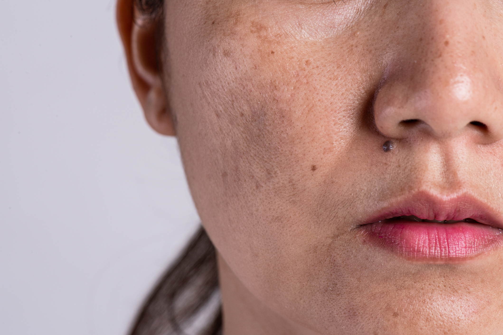
Melasma causes flat brownish patches with irregular borders particularly affecting the cheeks, temples and forehead, upper lip and nose. It can also affect the jawline and less commonly the forearms and upper chest. Facial involvement is generally symmetrical i.e. the same areas are involved on both the left and right side of the face.
The colour of healthy skin is largely determined by a pigment known as melanin that is produced by a specialized type of cell called a melanocyte. These cells are located in the surface layer of the skin and increased production by melanin plays a key role in the development of melasma. This tendency often runs in the family and is exacerbated by a number of triggers including hormonal factors and light exposure.
If you would like to learn more about what is understood about the mechanisms of melasma I recommend the following articles: Lee et al and Kwon et al.
Melanin pigment is produced within melanocytes from a naturally occuring amino acid (L-tyrosine) through a series of chemical reactions. The most important of these conversion steps requires the function of a protein "enzyme" within the cell called tyrosinase. Many of the treatments for melasma block the function of tyrosinase.
Melasma will often run in the family and this reflects a genetic predisposition for melanocytes to overproduce melanin in response to stimuli such as sunlight exposure.
Melasma is triggered by hormonal factors, particularly oestrogen. It will often worsen during pregnancy due to high oestrogen levels and for this reason it is also referred to as the "mask of pregnancy". Oestrogens in the combined oral contraceptive pill can also trigger melasma.
The role of ultraviolet (UV) light (present in sunlight) in causing melasma is well established. This is why melasma tends to worsen in the summer and to improve in winter months. This also explains why certain sites that receive higher levels of sunlight exposure - such as the cheeks, temples and forehead - are more frequently affected. For this reason, sun avoidance and the assiduous daily use of sun protection throughout the year is critical.
In recent years studies have revealed that in addition to ultraviolet light (UV), intense blue light causes pigmentation. For example Mahmoud et al showed that irradiating the back of volunteers with darker skin types with either blue light or ultraviolet light resulted in pigmentation. Regazzeti et al identified the mechanism of this (a protein, Opsin-3, that is present within cells and senses light).
Sunscreens were originally developed to stop sun burn, to protect from skin cancer and to protect against skin aging. All of these are mediated by ultraviolet light - specifically UVA and UVB. Most modern sunscreens are very effective at blocking ultraviolet light, however not all will block blue light. Castanedo-Cazares et al compared the effects of sunscreens that blocked UV light alone to those that blocked UV and visible (blue) light in 68 patients. They found that blocking blue light in addition ot ultraviolet improved the effects of depigmenting agents in treating melasma. It is therefore important to choose a sunscreen that blocks both UV light and blue light.
The skin is composed of a thin outer layer - the epidermis - that acts as a barrier and a deeper tough fibrous layer - the dermis that give strength to the skin. Melasma can cause pigmentation in either the epidermis, the dermis or both. The layer that is affected determines the appearance of the pigmentation and also has implications for treatment.
I cannot overemphasize how important sun protection is in treating and preventing melasma. As I have discussed above, melasma is caused not only by ultraviolet light - which almost all modern sunscreens will block effectively - but also by blue light. Blocking blue light is critical in the treatment of melasma and requires either an iron oxide based sunscreen or a chemical sunscreen with enhanced spectral coverage.
There are many effectice sunscreens available. I personally like products from the Anthelios XL and Heliocare 360 ranges. No sunscreen offers complete protection and therefore sun avoidance i.e. minimizing time spent in the sun particularly on sunny days is also important.
A range of over the counter topical products claim to improve pigmentation by reducing the production of melanin pigment.
Cysteamine cream is an emerging treatment option for melasma that is available without prescription. Lima et al treated 100 women with either depigmenting agents or topical 5% cysteamine cream. They found comparable improvements in pigmentation. Nguyen et al also looked at the efficacy of topical cysteamine in comparison with cysteamine in 20 patients with similar findings.
As discussed below oral antifibrinolytic agents are emerging as an effective option for melasma. Antifibrinolytic agents have also been used as a topical (cream) treatment. El-Husseiny et al treated 100 patients with topical antifibrinolytic agents on one side of the face and tyrosinase inhibitors on the other side. They found the efficacy of topical antifibrinolytic agents to be comparable to tyrosinease inhibitors.
The evidence supporting efficacy of these products is variable and they may be more suited to a maintenance regimen once pigmentation has been treated.
Relatively mild melasma will often improve with careful daily use of sunscreen, sun avoidance and non-prescription topical treatments. Patients will generally come to me when these measures have not proven effective. The first thing that I will do is to ensure that the diagnosis is correct and that there are not other co-existing causes for pigmentation.
Whilst diagnosis is generally straightforward, a number of other conditions can appear similar to melasma:
Facial pigmentation can be complex resulting from multiple causes. Where appearances are atypical or where there is failure to respond to standard treatments a skin biopsy can be considered.
Since the 1990s there has been good evidence that topical retinoids, such as tretinoin, improve melasma. Griffiths et al performed a randomized trial in 38 women. They found 13 (68%) of 19 patients treated with topical retinoids improved compared with 1 (5%) of 19 in the control group. Topical retinoids are often included along with tyrosinase inhibitors in combination treatments and are also used on their own as part of a mainenance routine.
Azelaic acid also has some benefit, it is less effective for melasma but unlike topical retinoids is safe for use in pregmancy.
Tyrosinase inhibitors, such as hydroquinone, are generally prescribed in combination products such as pigmanorm and reduce the synthesis of pigment (melanin) by melanocytes (pigment producing cells) by impairing the function of tyrosinase. They are often supplied in combination with a retinoid and mild topical steroid to enhance the effects and reduce the risks of an inflammatory reaction.
Tyrosinase inhibitors and combination products are one of the most effective treatments for melasma. The vast majority of my patients find topical tyrosinase inhibitors to be a safe and effective treatment, however as for all medicines they do have the potential for side effects. You can learn more about tyrosinase inhibitors at the following link:-
Tranexamic acid is an anntifibrinolytic agent and was originally developed for the treatment of major bleeding and is also commonly used as a treatment for heavy periods. In recent years evidence has emerged that oral antifibrinolytic agents can be an effective treatment for melasma. Lee et al analysed 561 patients treated with oral antifibrinolytic agents for melasma. They observed an improvement in 89.7% of patients usually within 2 months.
The mechanism by which antifibrinolytic agents improve pigmentation is poorly understood - it has been proposed that they reduce melanin synthesis and blood vessel formation in the skin.
Like many treatments used in dermatology, antifibrinolytic agents are used off licence for melasma - this means that it has not been formally approved for treatment of this condition. They are generally well tolerated, however as for any medicine there is a potential for side effects. Of these, the most important is the risk of blood clots. In otherwise healthy women this risk is very low. In the study of Lee et al, one patient out of 561 developed a deep vein thrombosis (blood clot in the leg). This patient was subsequently found to have a genetic disorder of blood clotting. For this reason, I would be very reluctant to prescribe oral antifibrinolytic agents where there is a personal or family history of blood clots. A genetic tendency to blood clots can be detected with blood tests and this can be considered when patients that are concerned or for patients taking oral antifibrinolytic agents on a long term basis.
Lasers and light treatments are generally reserved for patients that have failed to respond to the medical treatments (creams or tablets) discussed above. They do not cure the underlying tendency to pigmentation, however they can help in reducing pigment that is present. All of these treatments carry a risk of recurrence or flare on stopping treatment and they should usually be combined with a medical treatment such as topical depigmenting agents to minimize this risk. Response to treatment is unpredictable and patients with darker skin types are at greater risk of skin inflammation which can cause post inflammatory hyperpigmentation.
Since response to laser and light treatments is unpredictable I will often advise that we treat a small test area to begin with to assess the degree of improvement and to ensure that there are not complications such as increased pigmentation or patchy loss of normal pigment. I will also advise measures to reduce the risk of post inflammatory hyperpigmentation - this may include topical steroids prior to laser treatment and topical tyrosinase inhibitors before and after laser treatment.
The principle of laser and light treatments is that light energy is delivered to the skin at a specific wavelength that causes damage to pigment producing cells (melanocytes). These are subsequently eliminated by the immune system. A limitation is that too much damage can lead to reactive pigmentation post-inflammatory uyperpigmentation.
Non ablative fractional lasers use a wavelength of light that causes little damage to the surface layer of the skin (epidermis), but causes microscopic columns of injury to the underlying dermis. This results in remodeling and collagen synthesis in the dermis. It is believed that the removal of melanocytes during this healing process is the mechanism underlying improvement in melasma. Lasers in this category most commonly have wavelengths of either 1540 or 1927 nanometers.
Tourlaki et al treated 76 patients who had previously failed to respond to topical tyrosinase inhibitors alone with 1540nm fractional non-ablative laser and tyrosinase inhibitors combination topical treatments. At 1 month following treatment marked (>75%) and moderate (51-75%) clearing of melasma was observed in 67% and 21% of cases, respectively.
Q-switched lasers produces a wavelength of light that targets the pigment-producing cells (melanocytes) within the skin. The duration of the pulse is very short enabling relatively selective targeting of melanocytes. They are very effective for treatment of other causes of pigmentation on the skin such as freckles and the hope was that they would also be effective for treatment of melasma, however results of early studies were disappointing with little improvement and often rebound worsening of melasma.
Disappointing results with Q-switched lasers used at normal power settings led to the concept of using these lasers at a much lower power "low fluence" but repeating treatments more frequently. Wattanakrai et al treated one half of the face in twenty-two patients in this manner. The side treated with laser achieved an average 92.5% improvement compared with 19.7% on the control side. However patchy color loss "mottled hypopigmentation" developed in 3 patients, 4 patients developed rebound pigmentation, and all patients had recurrence of melasma. Subsequent studies are largely consistent with these results showing improvement in a proportion of patients but significant risks of post-inflammatory hyperpigmentation, mottled pigment loss (hypopigmentation) and a high rate of recurrence of melasma. For this reason I generally reserve Q-switched laser treatments for patients that have failed to respond to other available treatments.
A newer development is Picosecond lasers which have had better results with lower risk. I do not offer this treatment, however it is available via other specialists in London.
A tendency to melasma is present lifelong and therefore intensive life-long protection from both ultraviolet and blue light is a critical part of a maintenance regimen. Topical retinoids and non prescription topical treatments also play a role here.
Melasma can be frustrating to treat, both for the patient and the doctor. It is slow to respond to treatment, especially if it has been present for a long time however with patience and diligent adherence to treatment good results can be achieved in most patients.
To book an in person consultation, enter your details below and my practice management team will contact you to schedule the appointment. Alternatively call 0203 389 6076 (calls are answered during working hours) or email: contact@drmagnuslynch.com.