Skin cancer checks are vital to detect skin cancer early, when treatment is most effective.
Early detection leads to higher survival rates: Almost 100% of people with melanoma skin cancer will survive for 5 years if the disease is caught at its earliest stage. [Cancer Research UK]
Melanoma is the fifth most common cancer in the UK. There are around 17,500 new melanoma skin cancer cases in the UK every year, that’s 48 every day. [Cancer Research UK]
Consultant Dermatologist. Mohs and reconstructive surgeon. Laser Surgeon.
Harley Street, The London Bridge Hospital, OneWelbeck and Guy's Hospital.
21 years experience. >12 years exclusively in Dermatology. Thousands of skin surgery procedures, a large proportion on the face.
Medicine at Cambridge University. PhD at Oxford University. Dermatology training and Mohs fellowship at world-renowned St John's Institute of Dermatology.
Featured in channel 5 TV's Skin A&E. Research leader King's College London. Regular contributor to national media.
“Excellent service with a very approachable and friendly manner. Very clear assessment and advice. Very happy with the appointment.”
“Dr Lynch was thorough and gave excellent explanations when diagnosing and the subsequent follow up care. I felt very reassured following the consultation.”
“The treatments provided were top-notch, and the follow-up care made me feel valued as a patient.”
The most dangerous form of skin cancer, Melanoma usually appears as a dark or irregular mole.
This generally appears as a rapidly growing lump or non-healing area. It can usually be cured by surgery but does have the potential to spread.
This appears as a slowly growing lesion that is often pearly in appearance. It almost never spreads but can cause significant tissue destruction.
If you have a mole or other skin lesion that has changed in appearance (colour, shape or size), have moles that bleed, itch, ooze or crust over, or have new moles, we strongly recommend you have them checked.
All forms of skin cancer are more common in individuals who have had increased levels of sun exposure or who have used sun beds. Skin cancer is also significantly more common in those with lighter skin types. Other risk factors for skin cancer include:
Firstly, Dr Lynch will invite you to share any specific concerns you may have, and take a detailed history including any relevant family history of skin cancer.
Lesions of particular concern will be examined with a special microscope called a dermatoscope.
If you would like a full skin check, a comprehensive examination will be performed starting from the head and neck and working through all body areas.
Dr Lynch will take dermoscopic photographs of moles that require monitoring. If you have a large number of moles Dr Lynch can arrange for you to have mole mapping photographs taken (this will require a separate visit and additional charges will apply).
If Dr Lynch is concerned about a skin lesion then he may recommend a skin biopsy to be taken.
Either a sample of the lesion or the entire lesion is removed under local anaesthetic for analysis. Depending upon the nature of a skin lesion it can be removed either by cutting and stitching to leave a straight line scar or by shaving parallel with the skin surface and I will discuss the different options with you. The lesion is then sent for analysis by an experienced pathologist with whom I have a close working relationship and results are typically available within 1-2 weeks.
Dr Lynch has advanced training in skin surgery and have performed thousands of skin biopsies so where this is required I can perform this with minimal discomfort and scarring.
If you have a number of irregular moles Dr Lynch may advise that you have mole mapping photographs performed. This enables me to monitor for any change in your moles over time.
The principle of mole mapping is that harmless moles either do not change in appearance or change very slowly over years whereas melanomas usually will change significantly in appearance over a number of months.
Mole mapping consists of photography of the skin surface. It serves as a photographic record of lesions on the skin for comparison in future consultations. The photography itself does not perform any analysis of the skin and does not replace a comprehensive examination of the skin by a dermatologist with a dermatoscope (microscope). For any moles that I am concerned about, I will take additional digital dermascopic photographs of the pigment pattern. This allows me to check for any subtle changes at a follow up appointment.
Even if you are having regular skin checks with a dermatologist it is important that you monitor your own skin at home. You should perform a full skin check every few months - this has the benefit that you learn what is normal on your own skin and learn to recognise any change.
An easy way to monitor for change is to take photographs of all of your skin (you will need help for some areas) and then repeat these photographs after a few weeks/months. If you notice any new skin lesions or any change in the appearance (size, shape, color) of existing skin lesions it is important that these are reviewed.
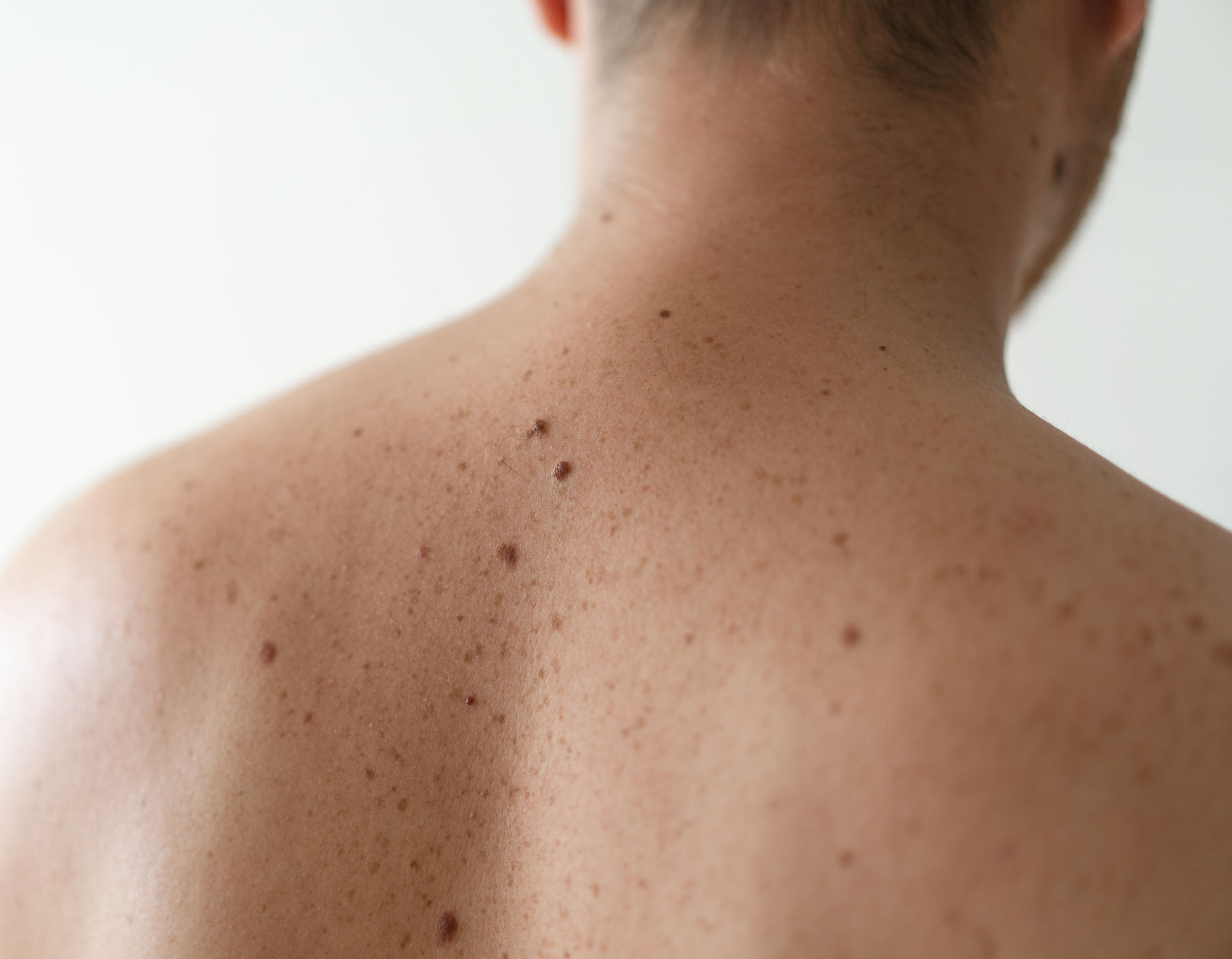
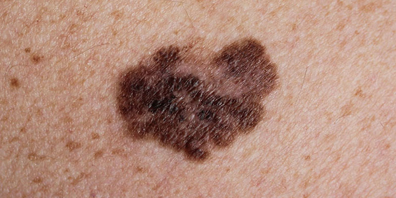
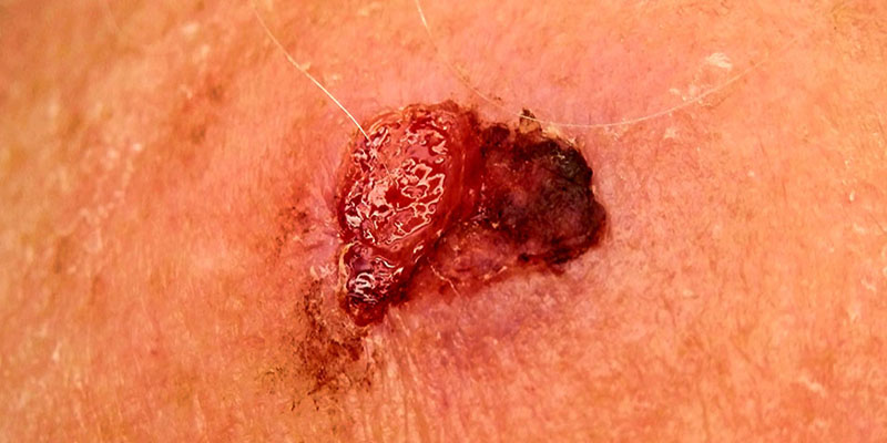
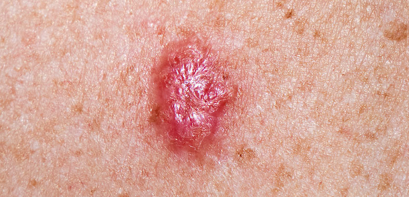
Dr Magnus Lynch is passionate about advancing our understanding of skin cancer through cutting-edge research. His work spans from pioneering spatial mapping of cell populations in skin cancers like basal cell carcinoma to investigating the potential of imaging technologies such as Optical Coherence Tomography, and understanding weaknesses in machine learning systems for skin cancer detection. His earlier research uncovered how spatial dynamics influence the behaviour of mutant cells in the skin, shedding light on the earliest stages of cancer development. He has also led a clinical trial exploring the potential for non-invasive diagnosis of melanoma through epigenetic profiling of the skin surface. Current work in the lab focuses on 3-dimensional cell culture models of skin cancer.
SELECTED SKIN CANCER RESEARCH PUBLICATIONS:
Ganier C, Mazin P, Herrera-Oropeza G, Du-Harpur X, Blakeley M, Gabriel J, Predeus AV, Cakir B, Prete M, Harun N, Darrigrand JF, Haiser A, Wyles S, Shaw T, Teichmann SA, Haniffa M, Watt FM, Lynch MD. Multiscale spatial mapping of cell populations across anatomical sites in healthy human skin and basal cell carcinoma. Proc Natl Acad Sci, 2024. Pubmed ID: 38165934
Lynch MD. Beyond the algorithm: Ethical Challenges in AI-Driven Skin Cancer Diagnosis. British Journal of Dermatology, 2024. DOI: 10.1093/bjd/ljae490
Hughes S, Srenathan H, Lynch MD (Joint senior author), Leeman H. Multi-center experience from tertiary skin cancer units on the role of Sentinel Lymph Node Biopsy in patients with pT1b melanoma. Clin Exp Dermatol, 2023. Pubmed ID: 38112606
Wan B, Ganier C, Du-Harpur X, Harun N, Watt F, Patalay R, Lynch MD. Applications and future directions for Optical Coherence Tomography in Dermatology. British Journal of Dermatology, 2021. Pubmed ID: 32974943
Du-Harpur X, Arthurs C, Ganier C, Woolf RT, Laftah Z, Lakhan MK, Salam A, Wan B, Watt FM, Luscombe NM, Lynch MD. Clinically-relevant vulnerabilities of deep machine learning systems for skin cancer diagnosis. Journal of Investigative Dermatology, 2020. Pubmed ID: 32931808
Lynch MD, Lynch CNS, Craythorne E, Liakath-Ali K, Mallipeddi R, Barker JN and Watt FM. Spatial constraints govern competition of mutant clones in human epidermis. Nature Communications, 2017. Pubmed ID: 29066762